

- Overview
- Recommended Products
Specification
model |
parameter |
another name |
application |
DOM-600 |
Six - gear five - times zoom system |
Root canal microscope, oral microscope |
For use in general stomatology,refined treatment,visualized treatment |
Product general information
Place of Origin: |
Sichuan Province, Ziyang City |
Brand Name: |
ZHOEK |
Model Number: |
DOM-600 |
Certification: |
Registration certificate, production license, business license, patent certificate |
Product business terms
MOQ: |
1 |
Price: |
According to model configuration |
Packaging Details: |
DOM-600: Base:720*720*300 Frame:1180*590*260 Head packaging:600*470*320
|
Delivery Time: |
Within 15 days |
Payment Terms: |
|
Supply Ability: |
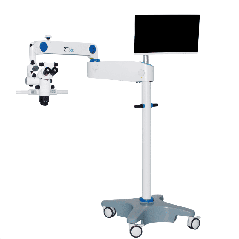
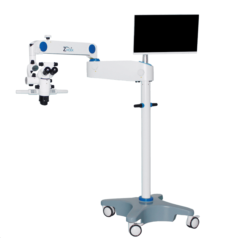
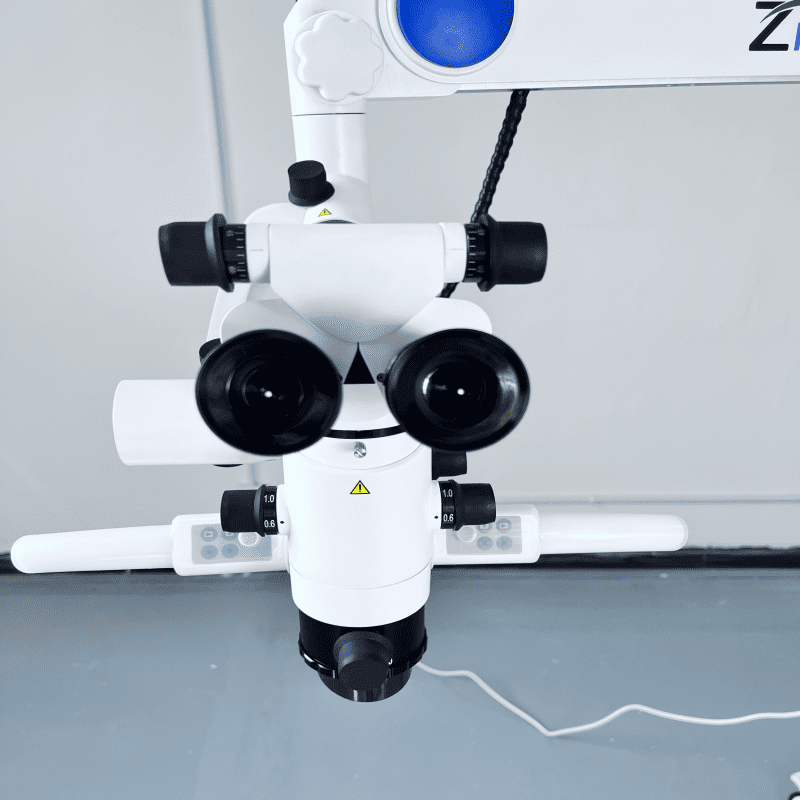
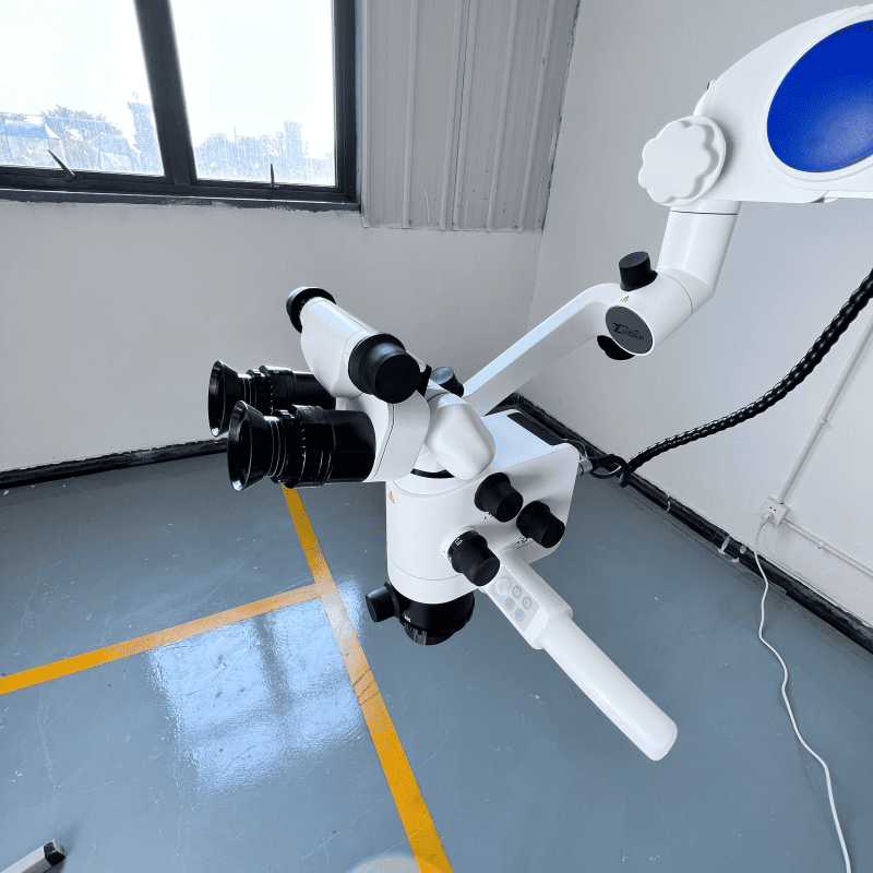
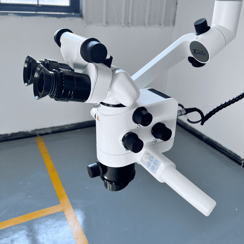
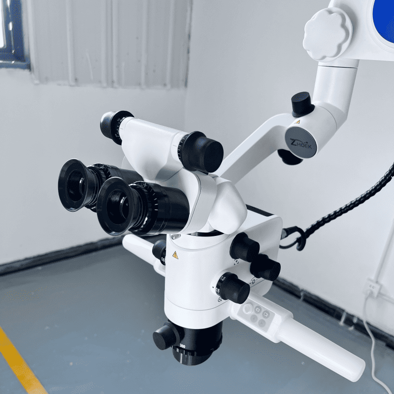
Description: Sichuan EST Medical Equipment Co., Ltd. has developed the DOM series of oral microscopes. The oral microscope is mainly used for oral examination and surgical treatment and is one of the essential devices in oral clinical practice. The company has developed four types of oral microscopes, and the series of products cover four - gear, five - gear, six - gear, and stepless zoom systems. They are equipped with high - tech features such as a fixed - focus fine - focus system, an LED uniform illumination system, and a balanced mechanical arm, which can provide a perfect oral solution. The company is committed to continuously exploring the clinical imaging needs of stomatology and developing characteristic imaging systems to meet the needs of oral clinical practice.
Alternative names for the product: Root canal microscope, oral microscope, surgical microscope
Core Specifications and Parameters:
DOM-600 Model Parameters |
|
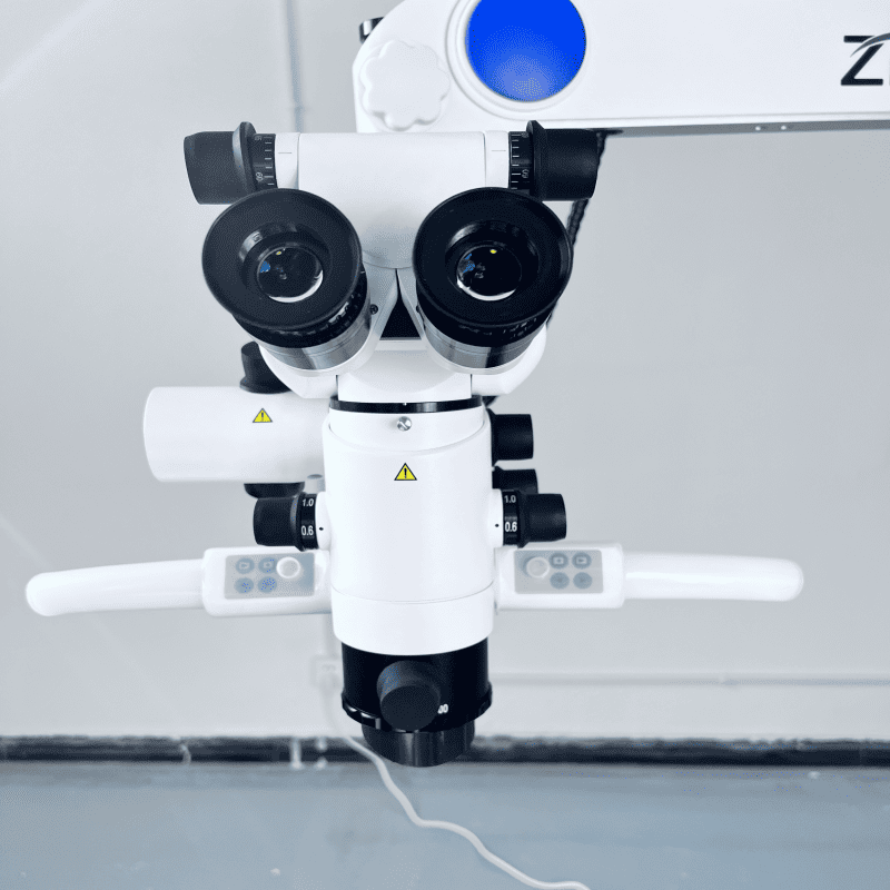
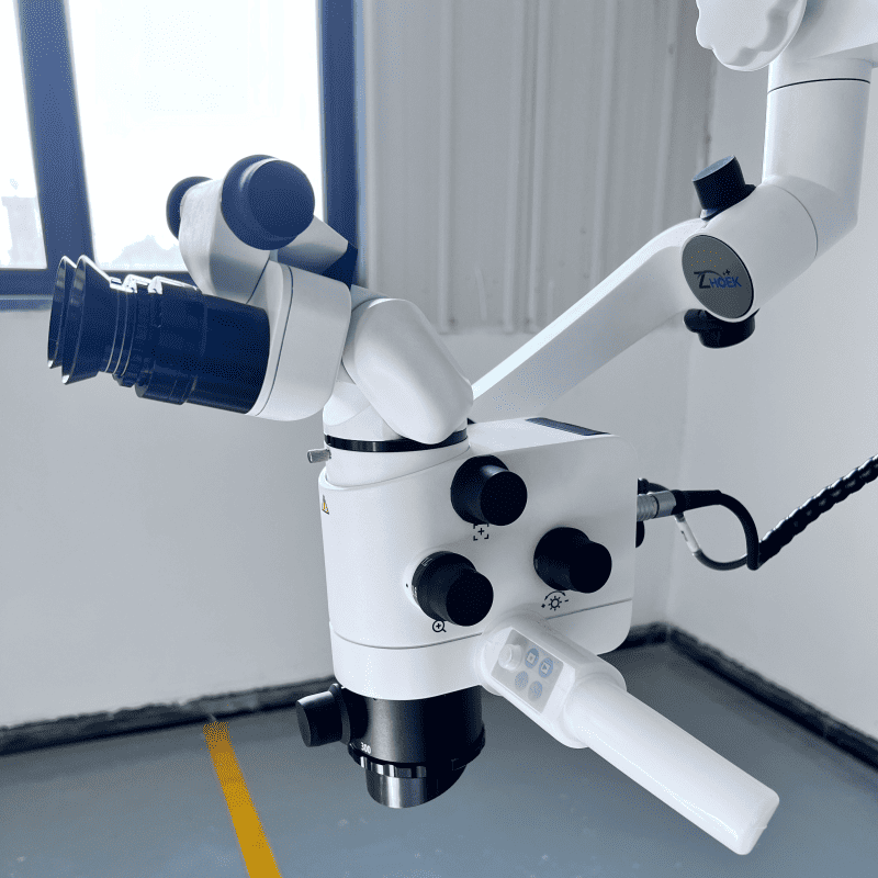
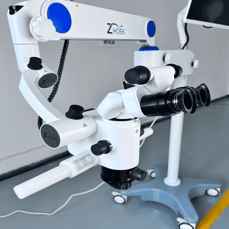
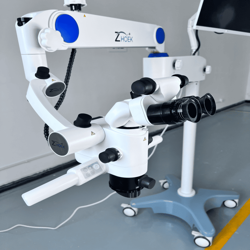
Optical System Interpupillary adjustment range: 55mm - 75mm, focusing accuracy ≤ 1mm; Eyepiece: Large - field (field - stop diameter 22mm) high - eye - point wide - angle eyepiece to obtain a larger field of view; Eyepiece magnification: 12.5X / 18B (optional 10X / 18B eyepiece); Refractive power adjustment range: ± 7D Binocular tube: 0 - 220° continuously adjustable angle binocular tube, meeting the operating posture of different doctors; Zoom method: 6 - gear five - times zoom system; Total magnification (when the large - object lens focal length f = 250mm): 3.6x, 6.4X, 12X, 18.4X, 26.5x, 6.4X; Zoom object lens: WD 180 - 300mm, focusing range 120mm; (optional super - zoom object lens: WD 200 - 450mm, focusing range 250mm) Field - of - view diameter (when the large - object lens focal length f = 250mm): 80mm, 68.2mm, 37.2mm, 14.9mm, 4.5mm, 68.2mm; Zoom factor (drum factor or coefficient): 0.4x, 0.6X, 1X, 1.6X, 2.5x, 0.6X; Illumination System Lighting source: Dual - channel top - grade medical LED light source, non - spherical uniform illumination technology, continuously adjustable brightness, service life ≥ 100,000h; Illuminance on the object surface (f = 250mm): ≥ 80,000LX; Illumination spot diameter: ≥ Φ85mm; Illumination color temperature: 6500K, close to natural light, true - to - life colors; Two types of color filters and light - spot adjustment modes: Orange, to prevent resin materials from curing prematurely; Green: to see tiny blood vessels and nerves under surgical blood environment; Single tooth, the field of view is more focused and not affected by artificial or natural light; (III) Frame System 1. Frame type: Standard pillar type (custom - made ceiling - mounted, wall - mounted, floor - fixed, desktop, iron plate, clamping type); 2. Damping balance hanging arm: Equipped with a 120° damping balance hanging wall balance adjustment microscope body, adjustable torque balance and damping, left and right deflection angle ± 45°, tilt angle ≥ 90°. When adding external cameras and other accessories, the gravity distribution can be readjusted to keep the microscope body in a smooth balance state; 3. Rotary arm: Length 830mm, rotation angle 270º, vertical movement 600 mm; 4. Horizontal arm: Length 450mm, rotation angle 360°; 5. Maximum net arm span: ≥ 1528mm; 6. Base size: 640mm × 640mm; (IV) Imaging System | | Built - in Imaging | | 1. Video frame rate: 1080P built - in module 19201080 @ 60fps (switchable to 960P, 720P); (optional 4K built - in module 38402160 @ 60fps (switchable to 2.7K, 1440P, 1080P, 960P, 720P)); | | 2. Optional stereoscopic beam splitter: A 45° optical extender integrated with the beam splitter, split - light ratio 2:8 / 5:5, pendulum system: Under the condition that the doctor's sitting posture remains unchanged, the binocular tube can keep the horizontal observation position while tilting left and right by 25° | | 3. Storage medium: 32 G USB drive (supporting 128G / 256G); | | 4. Playback function: Instant playback of the captured image files on the machine; | | 5. Supported shooting modes: JPG \ RAW (image format), MP4 (video); | | 6. Control method: One - key control handle, one - key photo and video recording; (optional: Wireless foot pedal, mobile phone, Android all - in - one machine and other terminals to achieve photo and video recording control, and can also install APP for remote medical treatment, remote teaching and other fields; ) | | 7. High - definition monitor and bracket: Full - HD equipped with a high - definition monitor, with an HDMI interface, the monitor bracket is used to stabilize the high - definition monitor and is installed on the microscope body;
|
|
Name and Specification of Goods Surgical microscope (floor - standing type) 0 - 220° adjustable angle binocular tube, precise interpupillary adjustment mechanism LED light source 6 - gear five - times zoom system Balanced damping hanging arm Zoom object lens F = 180mm - 300mm (optional: F = 200mm - 450mm super - zoom object lens) Imaging System: Built - in 1080p camera system (optional: 4K imaging system) 2. Video and photo control handle 3. High - definition 1080P monitor (optional: 4K monitor) 4. Monitor bracket 5. (Optional: Two - in - one stereoscopic beam splitter with tilting and extending functions) 6. (Optional: Built - in wireless foot pedal, wireless photo and video recording with foot control)
|
Applications: Dental pulp and periodontal disease treatment, periodontal and implantology, oral and maxillofacial surgery, aesthetic restoration, etc., and can also be used for teaching and research applications.